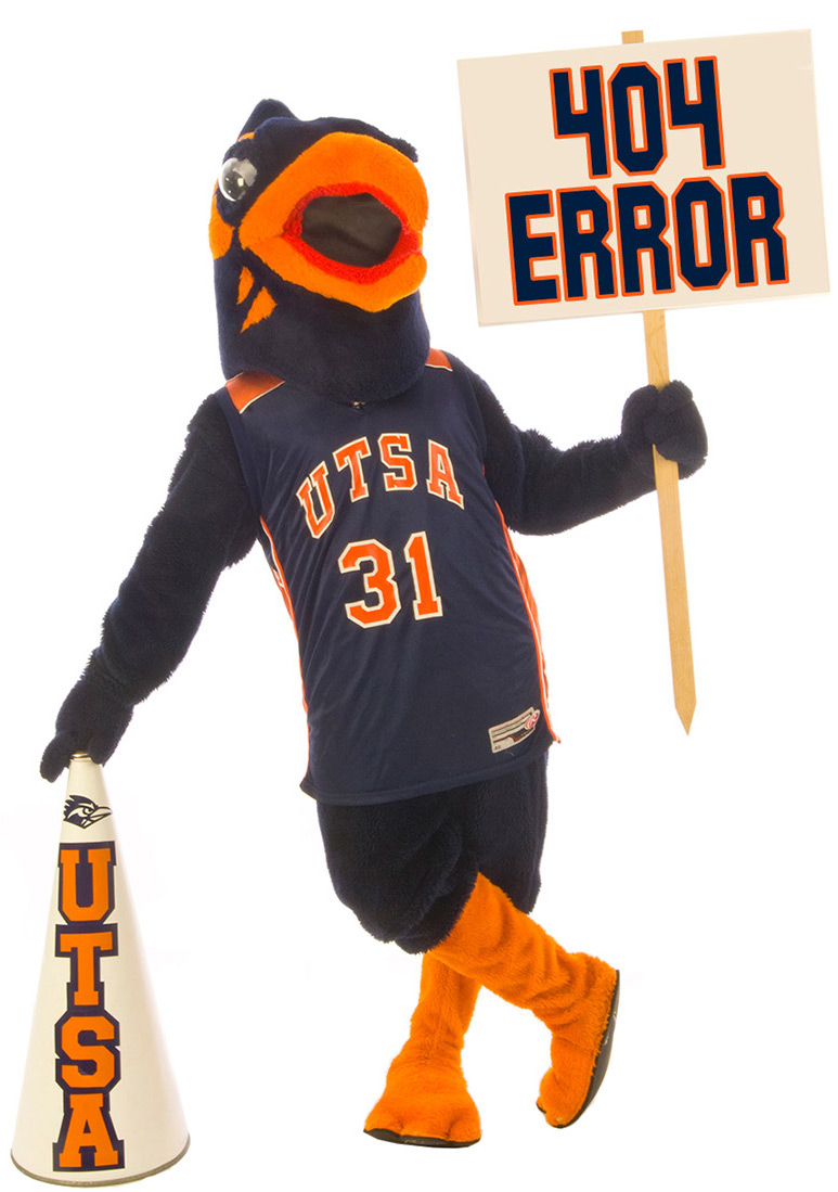
Quantitative, three-dimensional analysis of granule cell dendrites in the rat dentate gyrus.
Brenda J. Claiborne, David G. Amaral, and Maxwell Cowan
The Salk Institute for Biological Studies
San Diego, California 92138
J Comp Neurol, 1990 Dec 8; 302(2):206-19


ABSTRACT

The three-dimensional organization of dentate granule cell dendritic trees has been quantitatively analyzed with the aid of a computerized microscope system. The dendrites were visualized by iontophoretic injection of horseradish peroxidase into individual granule cells in the in vitro hippocampal slice preparation. Selection criteria insured that the analyzed cells were completely stained and that only neurons with two or fewer cut dendrites in the distal portion of the molecular layer were analyzed. Twenty-nine of the 48 sampled granule cells had no cut dendrites. The granule cells had between one and four primary dendrites. Granule cell dendritic branches were covered with spines and most extended to the hippocampal fissure or pial surface. The mean total dendritic length was 3,221 microns with a range from 2,324 microns to 4,582 microns. The dendrites formed an elliptical plexus with the transverse spread averaging 325 microns and the spread in the septotemporal axis averaging 176 microns. On individual neurons, the maximum branch order ranged from four to eight and the number of dendritic segments ranged from 22 to 40. Approximately 63% of the dendritic branch points occurred in a zone that included the granule cell layer and the inner one-third of the molecular layer. The dendritic tree was organized so that, on average, 30% of the length was in the granule cell layer and proximal third of the molecular layer, 30% was in the middle third, and 40% was in the distal third. Comparisons were made between the dendrites of granule cells in the suprapyramidal and infrapyramidal blades of the dentate gyrus. Suprapyramidal cells had a significantly greater total dendritic length than infrapyramidal cells, their transverse spread was higher, and they had a greater number of dendritic segments. When neurons in the suprapyramidal blade were further subdivided on the basis of somal position within the depth of the cell body layer, superficial neurons were found to have a greater number of primary dendrites, more elliptical trees, and larger transverse spreads of their dendrites. There were no significant differences in dendritic segment number or total dendritic length between superficial and deep cells.

Figure 1

Photomicrographs of HRP-filled granule cells. A: Low-magnification photomicrograph of the dentate gyrus in a hippocampal slice preparation. A filled granule cell is present in the infrapyramidal blade (open arrow); its cell body is in the granule cell layer (GL) and its dendrites extend into the overlying molecular layer (ML). PL; pyramidal cell layer. B: A higher-magnification photomicrograph of an HRP-filled granule cell located in the suprapyramidal blade. Only a portion of the dendritic tree is in focus. Stained red blood cells can be seen scattered throughout the slice. The axon (a mossy fiber) exits from the base of the soma (asterisk). C: A higher-magnification photomicrograph of a portion of the dendritic tree of a granule cell showing dendritic spines of several sizes and shapes (arrows). Scale bars: A = 100 mm; B = 50 mm; C = 10mm.

Figure 2

Computer-generated plots of reconstructions of two HRP-filled granule cells from the suprapyramidal blade. Each neuron is viewed from three different spatial angles. The neuron shown in the top panel (a) did not have any cut dendrites while the neuron illustrated in the lower panel (b) had two cut dendrites (marked by asterisks) in the distal part of the molecular layer. In a1 and b1, the cells are viewed in the transverse plane of the dentate gyrus (i.e. in the same orientation as they were entered into the computer system). The top panel is used to illustrate nomenclature for branch order; 1st-, 2nd-, and 3rd-order dendritic branches are indicated, as are the primary dendrite and a dendritic segment. For the views in a2 and b2, the cells were rotated 90° about their vertical axis, illustrating their dendritic spreads in the longitudinal plane of the dentate gyrus. In a3 and b3, the cells are viewed from above (i.e., from the pial surface).

Figure 3

Computer-generated dendritic tree diagrams from six different granule cells, three of which were located in the suprapyramidal blade (A-C) and three in the infrapyramidal blade (D - F). The three dimensional lengths (in mm) of each segment are given. Scale bar = 100 mm.

Figure 4

Computer-generated plots of reconstructions of the dendritic the total dendritic trees from granule cells located at various positions around the transverse axis of the dentate gyrus. Each neuron was from a different animal. The total dendritic length of each neuron is indicated. Note that the total dendritic lengths of the neurons in the suprapyramidal blade are greater than those in the infrapyramidal blade. CA3 : field CA3 of hippocampus.

Figure 5

Micrographs showing multiple labeling of granule cells from a single HRP injection. In A and C, two neurons were filled, while in B and D, three neurons were labeled. Close examination of each of these preparations indicates that a dendrite from one filled cell runs directly over (or under) the filled soma of another cell; we assume that it is at such sites that the HRP labeling has spread from the injection and into the adjoining neuron(s). This is especially clear in panel C where one of the dendrites from the labeled cell at the bottom can be seen to course over the filled soma of the top cell (open arrow). In many of the micrographs, an axon (asterisk) can be seen exiting from the granule cell body. Scale bar = 100 mm.


TABLE 1. Dendritic Parameters for Entire Population of Granule Cells1


Mean

(n = 48)

Range

Primary dendrites

1.9 ± .2

(1 - 4)

Dendritic segments

29 ± 1

(22 - 40)

Maximum Branch Order

5.7 ± .1

(4 - 8)

Transverse spread (m m)

325 ± 11

(186 - 518)

Longitudinal spread (m m)

176 ± 6

(87 - 286)

Total dendritic length (m m)

3,221 ± 78

(2,324 - 4,582)

Tree shape2

.56 ± .03

(.14 - .98)

Somal width (m m)

10.3 ± .3

(6 - 15)

Somal length (m m)

18.6 ± .5

(15 - 30)

1Values are means ± S.E.M.

2See text for method of determining tree shape.


TABLE 2. Percentages of Branch Points and Total Dendritic Length in Thirds of the Molecular Layer1

Thirds

Percent branch points

Percent total dendritic length

Proximal2

63 ± 2

(31 - 88)

30 ± 1

(11 - 55)

Middle

27 ± 2

(6 - 50)

30 ± 1

(23 - 41)

Distal

10 ± 1

(0 - 28)

40 ± 1

(20 - 52)

1Values are means ± S.E.M. All 48 neurons were included

2Branch points and dendritic length in the cell layer were included in the proximal third percentages.


TABLE 3. Dendritic Parameters: A Comparison of Suprapyramidal Vs.

Infrapyramidal Granule Cells1


Suprapryamidal

(n = 30)

Infrapyramidal

(n = 18)

P

Primary dendrites

2.1 ± .2

(1 - 4)

1.6 ± .2

(1 - 4)

¾

Dendritic segments

31 ± 1

(24 - 39)

27 ± 1

(22 - 40)

*

Maximum Branch Order

5.8 ± .2

(4 - 8)

5.5 ± .2

(4 - 7)

¾

Transverse spread (m m)

347 ± 14

(186 - 518)

288 ± 16

(187 - 445)

*

Longitudinal spread (m m)

182 ± 8

(87 - 286)

166 ± 6

(132 - 217)

¾

Total dendritic length (m m)

3,478 ± 88

(2,500 - 4,583)

2,793 ± 74

(2,324 - 3,450)

*

Tree shape2

.53 ± .04

(.14 - .98)

.59 ± .04

(.31 - .98)

¾

Somal width (m m)

10.4 ± .4

(8 - 15)

10.1 ± .4

(6 - 13)

¾

Somal length (m m)

19.3 ± .6

(15 - 30)

17.6 ± .6

(15 - 25)

¾

Molecular layer width (m m)

254 ± 7

(182 - 341)

240 ± 4

(189 - 274)

¾

1Values are means ± S.E.M. Ranges are in parenthesis.

2See text for method of determining tree shape.

* Significant at P < .05.


TABLE 4. Dendritic Parameters of Suprapyramidal Granule Cells: A Comparison of Superficial Vs. Deep Neurons1


Superficial2

(n = 19)

Deep

(n = 11)

P

Primary dendrites

2.4 ± .3

(1 – 4)

1.5 ± .2

(1 – 3)

*

Dendritic segments

31 ± 1

(24 – 39)

30 ± 1

(24 – 37)

¾

Maximum branch order

5.5 ± .2

(4 – 7)

6.4 ± .3

(5 – 8)

*

Transverse spread (m m)

378 ± 16

(255 – 518)

293 ± 16

(186 – 369)

*

Longitudinal spread (m m)

171 ± 11

(87 – 253)

200 ± 12

(148 – 286)

¾

Total dendritic length (m m)

3,484 ± 130

(2,500 – 4,582)

3,468 ± 92

(3,062 – 4,127)

¾

Tree shape3

.46 ± .04

(.14 - .78)

.67 ± .05

(.42 - .98)

*

1 Values are means ± S.E.M. Ranges are shown in parenthesis.

2 Superficial neurons had somata in the top half of the granule cell layer, whereas deep neurons had somata in the bottom half.

3 See text for method used to determine tree shape.

* Significant at P < .05.


TABLE 5. Dendritic Parameters of Infrapyramidal Granule Cells: A Comparison of Superficial Vs. Deep Neurons1


Superficial2

(n = 12)

Deep

(n = 6)

P

Primary dendrites

1.7 ± .3

(1 – 4)

1.3 ± .2

(1 – 2)

¾

Dendritic segments

28 ± 1

(22 – 40)

25 ± 1

(23 – 27)

¾

Maximum branch order

5.5 ± .3

(4 – 7)

5.5 ± .3

(5 – 7)

¾

Transverse spread (m m)

311 ± 17

(222 – 445)

244 ± 26

(187 – 321)

*

Longitudinal spread (m m)

170 ± 8

(132 – 217)

157 ± 8

(132 – 188)

¾

Total dendritic length (m m)

2,875 ± 95

(2,364 – 3,450)

2,629 ± 86

(2,324 – 2,857)

¾

Tree shape3

.55 ± .04

(.31 - .74)

.69 ± .09

(.42 - .98)

¾

1 Values are means ± S.E.M. Ranges are shown in parenthesis.

2 Superficial neurons had somata in the top half of the granule cell layer, whereas deep neurons had somata in the bottom half.

3 See text for method used to determine tree shape.

* Significant at P < .05.


TABLE 6. Dendritic Parameters: A Comparison of Adjacent Granule Cells


Suprapyramidal

a1

b

Infrapyramidal

a1

b

Primary dendrites

2

1

3

1

Dendritic segments

32

27

25

25

Maximum branch order

6

6

4

5

Transverse spread (m m)

420

389

332

252

Longitudinal spread (m m)

87

121

184

196

Total length (m m)

3,155

3,102

2,835

3,187

Tree shape2

.14

.31

.55

.73

1 Neurons labeled "a" and "b" were adjacent to one another. The cell bodies of all neurons were located in the superficial half of each cell layer. None of the dendrites were cut

2 See text for method used to determine tree shape.