Gallery of Cool Images

Rat Spermatogenesis
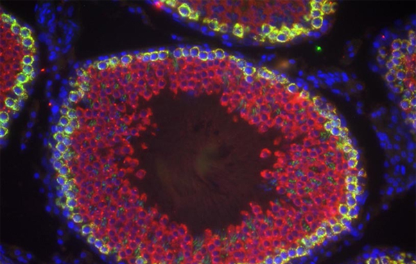
This image shows immunofluorescent staining of tissue sections from an adult Sprague-Dawley rat testis for the pan germ cell marker Vasa (Ddx4, Red) and the piRNA interacting protein Piwil2 (green). Colocalization of these two cytoplasmic proteins is shown in yellow. Stained sections were counterstained with DAPI (blue) to mark nuclei.
image taken using a SPOT-RT slider camera on a Nikon Eclipse E600 upright microscope in the laboratory of Dr. Kyle Orwig in the Magee-Womens Research Institute at the University of Pittsburgh

Rat Embryonic Testis
This image shows immunofluorescent staining of tissue sections from embryonic Sprague-Dawley rat testes for the pan germ cell marker Vasa (Ddx4, Red) and the marker of proliferation Ki67 (green). Stained sections were counterstained with DAPI (blue) to mark nuclei. Absence of Ki67 staining in VASA+ prospermatogonia inducates these cells are in mitotic arrest period of embryonic testis development.
image taken using a SPOT-RT slider camera on a Nikon Eclipse E600 upright microscope in the laboratory of Dr. Kyle Orwig in the Magee-Womens Research Institute at the University of Pittsburgh

Prepubertal Rhesus Testis Cells
Shown is a phase-contrast brightfield image of a suspension of testis cells from a prepuberal rhesus macaque generated by two-step enzymatic disruption of the testis (Collagenase IV then Trypsin + DNaseI). Somatic cells are small, round, and have a whitish appearance. Spermatogonia (Adark and Apale) are larger and have a brown-organge color.
image taken using a SPOT-RT slider camera on a Zeiss AxioVert40-CFL inverted microscope in the laboratory of Dr. Kyle Orwig in the Magee-Womens Research Institute at the University of Pittsburgh

Diatom Christmas Tree
This image shows a prepared slide containing diatoms arranged in the shape of a Christmas Tree (prepared by Klaus Kemp).
image taken using an AxioCam MRc5 camera on a AxioVert40 CFL inverted microscope with a 10X Objective and Differential Interference Contrast

Immunoflourecence in SSC Culture
This image shows immunofluorescent staining of DBA/2 mouse SSC culture stained for the SSC marker Plzf (red) and another transcription factor expressed in SSCs, Sall4 (green). Stained cells were counterstained with Hoechst 33342 (blue) to mark nuclei. Note the overlap between all three stains is displayed as white coloring in nuclei of labeled cells.
image taken using an AxioCam MRc5 camera on a Zeiss inverted AxioVert40 CRL microscope; image credit to Kazadi Mutoji

W/Wv Transplant Recipient Testis
Shown is an image of a cross-section through a W/Wv mouse testis two months after being transplanted with cultured SSCs from the DBA/2 mouse strain. This recipient mouse is genetically sterile due to a mutation in the cKIT receptor which thereby prevents spermatogonial differentiation. Thus, any spermatogenesis must have arisen from transplanted cells.
mosaic stitched image composed of 70 individual tiled images acquired at 20X magnification with an AxioImager M1 microscope using an ICc1 camera

Microtubule Tips
A fusion protein between the EB3 protein, which labels microtubule tips, and the mEmerald fluorescent protein expressed in PK1 cells (pig kidney epithelial cells).
image acquired on an AxioObserver inverted microscope with an Apotome device and ERm camera; shown is one image of several from a time-lapse image series