
Feature
Think Small
UTSA enters the nano race with the addition of a revolutionary new microscope
What tool promises to advance research in an untold range of subjects, from cancer therapy to solar panels, electronics to archaeology?
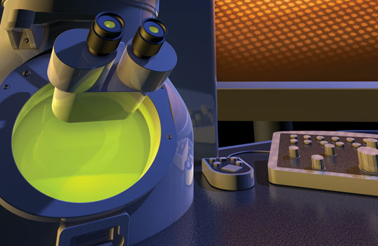
Illustration by Stephen Durke
This isn’t a trick question or even a riddle, but the answer does boggle the mind: This fall, a microscope with the ability to show atoms more clearly than ever will arrive at The University of Texas at San Antonio. When it does, it will be just the second of its kind in the world, according to the company that makes it. The other sits in Japan, in the factory of manufacturer JEOL, a global supplier of scientific instruments that specializes in electron microscopes.
The Robert J. Kleberg, Jr. and Helen C. Kleberg Foundation this winter gave UTSA $1.2 million, the final amount needed to purchase the second-generation aberration-corrected electron microscope nicknamed “Helenita” for foundation president Helen Kleberg Groves. UTSA physics and astronomy department chair Miguel J. Yacaman, a renowned electron microscopist and nanotechnology researcher, says the capabilities of this microscope, the best aberration-corrected microscope at a U.S. university, are legion. What makes the instrument so useful is its improved resolution and its ability to correct distortion, a problem Yacaman compares to the skewed effect of fun-house mirrors at a carnival.
“What you want is that your microscope doesn’t distort the real image,” he says. It packs additional features that make researchers swoon, such as the ability to analyze the chemical makeup of a sample and to reconstruct two-dimensional samples into three-dimensional images.
Three other powerful microscopes arrived in the Advanced Microscopy and Nanotechnology Lab on the Main Campus last year. The trio—a scanning electron microscope that shows three-dimensional images at high resolution, and two atomic force scanning probe microscopes that can measure the surfaces of nanoparticles—were funded with an $822,000 gift, also from the Kleberg Foundation. These new tools already have exponentially grown the lab’s sophistication and capabilities and offer their own specialized functions.
“There was no high resolution machine at UTSA,” Yacaman says. “There was no chance to look at nanoparticles on a scanning microscope. … We didn’t have atomic force microscopes before in the university.”
The aberration-corrected microscope “is going to be the first one of this kind in the United States, and it will allow researchers in many fields … to do work at the very high resolution level,” he explains. When it joins the lab, including the three other Kleberg-funded microscopes, the new microscope “will make [the lab] one of the most important microscopy facilities in the world,” he says.
And researchers in a host of fields, including materials science, chemistry, biology, industry and pathology, will be able to take part.
It’s a small world
It can be hard for the non-nano expert to fathom the tiny particles that are the bread and butter of researchers in this field. A nanometer is a billionth of a meter. Still fuzzy? A strand of human hair is about 20,000 nanometers in diameter. Fingernails grow one nanometer per second.
The ability to see small has evolved over the centuries, from the advent of optical microscopes in the 17th century that use light to make small things viewable, to the development of electron microscopes before World War II. Light has its limitations and can only go so far in resolution, but electrons, with their shorter wavelengths, go much further. Electron microscopes continued to improve over the years as well. But at the level of this new microscope, the potential for new discoveries is enormous, says Yacaman.
“Once you can see the atoms, you can learn a lot about how the matter is formed,” he says. “It’s a whole new ballgame.”
Just what kind of ballgame? Yacaman refers to the Hubble Space Telescope’s launch in 1990 as an example of a whole new world opened up. With the detailed and frequent images it provides, the Hubble has advanced our understanding of the universe, including the danger of cometary impacts, the evolution of galaxies and details of stellar death, according to the Space Telescope Science Institute.
“When they sent the Hubble to space, the number of discoveries that came from the Hubble were enormous,” he says. “So we expect with this microscope to have tremendous discoveries in the nano world.”
The Hubble telescope provides another example of the benefits of aberration correction, explains Ulrich Dahmen, director of the National Center for Electron Microscopy at the Lawrence Berkeley National Laboratory in Berkeley, Calif. The Hubble experienced its own aberration that required a space launch to correct, he explains. And the difference between the images from before and after that fix was obvious.
“What you see is a very clear difference in … how clearly you see the stars in the galaxy,” he says.
Under the microscope
Cancer research stands to benefit tremendously from the new technology. Researchers hope to design localized treatments that target cancer cells without causing damage to surrounding healthy tissue, as happens with conventional radiation treatment. Other applications—and there are many—include finding a substitute for pricey silicon crystals that can be used to make solar panels more efficient and cheaper, as well as developing better armor for military vehicles and creating improved antibacterials. Nanoparticles of silver already are used as an antibacterial in products, but Yacaman says there is more to be learned about them.
“Nanoparticles is one of the great ways to fight bacteria, but then of course we have to check on the negative effects it might have,” Yacaman says. “We need to design better materials for all kinds of applications, and one of the ways to do it is we have to know the atomic structure to really design a new material.”
College of Sciences Dean George Perry is planning to use the aberration-corrected microscope in his own research on oxidative stress and Alzheimer’s disease. Having such an instrument on campus is going to attract high-quality faculty whose research interests dovetail with the microscope’s features, he says.
“It will make it much easier for us to recruit top scientists,” he says. “It offers such resolution that it’s sort of way above what anyone can imagine.”
Gaining a tool as sophisticated as the aberration-corrected electron microscope at a university is a notable achievement, says Dahmen of the Berkeley National Lab.
“There’s no better way of illustrating your commitment to excellence in research,” he says. “A world-class machine like that shows you are willing to provide the support and infrastructure to give the best tools to your faculty.”
The microscope is making its debut at UTSA because of Yacaman’s reputation and longstanding relationship with the manufacturer, as well as the interest and support of the university, colleagues agree.
“Professor Yacaman is a world-class microscopist,” says Donald Paul, professor of chemical engineering and director of the Texas Materials Institute at the University of Texas at Austin. “This is going to be a very important tool for UTSA,” he continues. “I can easily envision there will be a number of people from Austin to use the microscope there.”
A former colleague and past collaborator of Yacaman’s, Paul notes a discovery of Yacaman’s in the mid-’90s that garnered widespread attention. He explained the longevity of the brilliant blue paint used by the Maya 1,000 years ago that had been baffling archaeologists and researchers for years. Yacaman found that the Maya blue, as it had come to be called, contained clay with nanoparticles of metal that kept the blue intact on ruins for centuries.
The new microscope will allow Yacaman to revisit Maya blue and perhaps resolve yet-unanswered questions.
“We are going to study Maya blue at ultrahigh resolution,” Yacaman says. “There is still some controversy on the subject that we will resolve with this machine. Discoveries like this will be common with the new machine.”
The microscope will be installed over a three-month period in the new engineering building, where the Advanced Microscopy and Nanotechnology Lab will be moving, and is expected to be fully functional by the end of the year. The plan is to make the microscope available to researchers around the country who will be able to access it remotely.
“They send a sample, we load the sample,” Yacaman says. “This instrument should be working 24 hours [a day]. The idea is to make it a Texas machine and eventually a USA machine.”
A magnet for students
The chance to work under Yacaman drew Alvaro Mayoral to UTSA. One of the Spanish postdoctoral fellow’s projects is making and analyzing metal nanoparticles that could be used in cancer and HIV research and semiconductors. He is using the new scanning electron microscope for his analysis.
“The fact that he is getting the new aberration-corrected microscope was a great bonus, although I would have come anyway even if he did not get that,” Mayoral says, adding, “I don’t think there’s a better place you can go” to pursue doctoral studies in microscopy or nanotechnology.
Before the trio of Kleberg-funded microscopes arrived last year, UTSA researchers were using a two-decades-old microscope with limited capabilities. “We could magnify to 60,000 times, and after that we were having trouble seeing things,” says David Olmos, facilities manager of the lab.
The scanning electron microscope has ramped that up to 2 million times magnification. Olmos compares the mighty progression of microscopy firepower at UTSA to trading up from a Volkswagen to a fine Maserati.
“At the nano level you have to have resolution because yeah, you can create all these little stars and particles of stuff that we’re going to be using for research, but unless you can look at them, what are we going to say? ‘Well, they’re in there … but we can’t see them?’ ” he says. “And the new microscope will take it to even a higher level.”
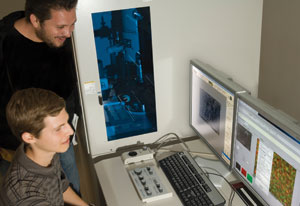
The lab also operates on a philosophy of empowering researchers, from undergraduates on up, to use equipment themselves, says Olmos. Already, some students have become adept at running the new equipment.
“Here it’s a user facility,” Olmos says. “That’s the way research is done.”
Rolando Valdez, a UTSA senior majoring in physics, is gratified by the role he is able to play in research using up-to-date equipment on his own. He recognizes the level of learning he is attaining.
“I think it’s a great place to be,” he says. “I feel like I’m a key player” in some of the projects.
Getting word out about these new resources at UTSA is important, says Bruce Nicholson, chair of the department of biochemistry at the University of Texas Health Science Center at San Antonio. Researchers will then consider how their own work might be enhanced and what previously out-of-reach questions might be answerable with the new technology.
A trio of powerful microscopes arrived in the Advanced Microscopy and Nanotechnology Lab on the Main Campus last year. They were funded with a gift from the Robert J. Kleberg, Jr. and Helen C. Kleberg Foundation.
UTHSCSA and UTSA already have been talking about collaborating on a drug discovery, development and delivery initiative that could tap into state funding for cancer research.
“Nanoparticles could be designed to emit heat and kill cancer cells,” Nicholson says, explaining the role of Yacaman’s lab in the collaboration. “It’s a directed missile instead of a huge nuclear bomb that will wipe out the whole city.”
To that end, UTSA assistant professor of physics Lorenzo Brancaleon is using the new scanning electron microscope and one of the new scanning probe microscopes in his research of small light-activated drugs and their effect on protein structures, one potential project in the collaboration between UTHSCSA and UTSA. He anticipates improved contrast in his samples using the aberration-corrected microscope.
“If someone can’t swallow because the tumor is in the esophagus and the food won’t go down,” the therapy can help, he says. “So they can eat again, so they can swallow again.”
The ability to see small has evolved over centuries to the point that scientists now can conduct their research and innovate solutions on a truly tiny scale. The nanoworld appears to be the new frontier, and UTSA is poised to push these explorations as never before.
“The support of the Kleberg Foundation to UTSA has permitted a quantum leap on the instrumentation for nanotechnology,” says Yacaman. “UTSA was far behind UT Austin or Rice on equipment for the physical sciences, and all of the sudden we are in the race.”
- Kate Hunger